|
HEISEI ANIMAL HOSPITAL
ULTRASONOGRAPHY |
◆HEART
A.Doppler Ultrasound
●BASIC EXAMINATION
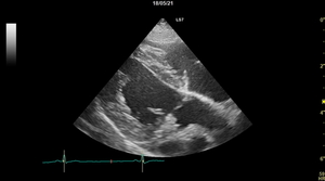
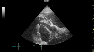
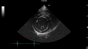
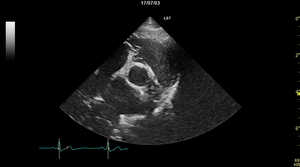
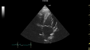
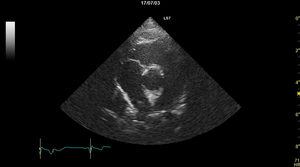
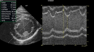
①heart morphology and movement of each cross-section
・long-axis four-chamber cross-section (right side) ・long-axis outflow tract cross-section (right side)
・short axis (chondral cord,papillary muscle ・short axis (heart) (basal level / right side)
level / right side)
・apical 4-chamber cross-section (left side) ・apical 5-chamber cross-section (left side)
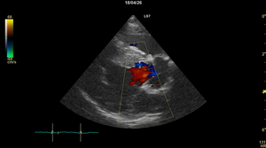
②Inspection of valve regurgitation by color doppler method【mitral valve・tricuspid valve・pulmonary valve・aortic valve】
etc…
◎diagnosis and staging of mitral regurgitation through
・confirmation of mitral regurgitation by color doppler method
・aortic valve regurgitation ・left atrial diameter measurement (ao/lv ratio)
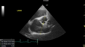
・left ventricular cavity wall measurement, inner diameter shortening rate measurement (FS%)
…etc.
③In the above test, cardiomyopathy(hypertrophy・restricted type・dilated type, etc.), stenotic lesions, and the prescence or absence of shunted blood vessels are also checked.
●INSPECTION OF +a
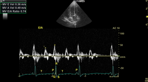 Left ventricular inflow blood flow velocity waveform inspection
E/A wave measurement
Can check the risk of left atrial pressure (pulmonary edema)
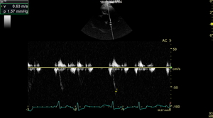
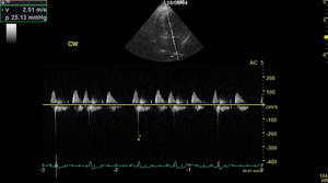 Reverse bloody velocity waveform measurement
For example, evaluating the severity of mitral regurgitation,
the left atrial pressure and left ventricular systolic state
If necessary, check for regurgitation of the tricuspid valve, pulmonary valve, and aortic valve
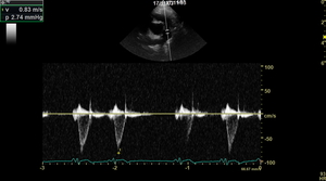 Blood flow waveform measurement
Aortic waveform shape and time measurement values,
tests the heart's contractile function, etc.
 Measurement of pulmonary arterial blood flow velocity waveform
Examine the presence and severity of pulmonary hypertension
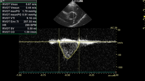 Stroke volume (SV), cardiac output (CO) measurement
Understand and inspect high and low output states
B.Tissue Doppler Inspection
↑Examine the condition of the heat muscle, red indicates muscle contraction and blue indicates muscle expansion.
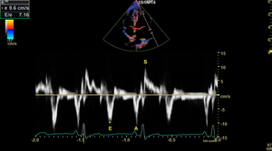 By measuring E/e ratio,
it is possible to diagnose the systolic dysfunction and diastolic dysfunction of the heart.
This image examines left atrial pressure, risk of pulmonary edema, left ventricular diastolic and systolic function
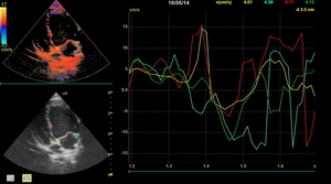 The movement of each part of the heart wall (myocardium) is measured,
to check the condition of the heart
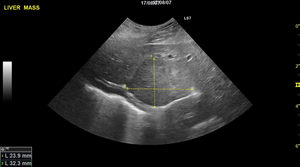
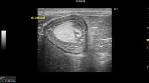
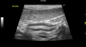
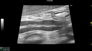
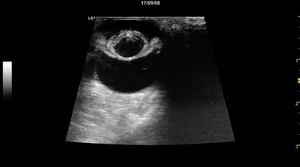
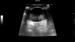
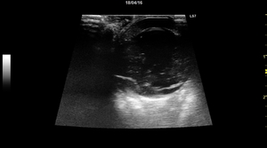
◆ABDOMINAL ORGANS
・Liver (liver cancer) ・Stomach
・Spleen ・Intestines
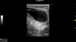
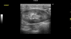
・Bladder (cystitis, stones) ・Kidney
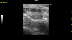
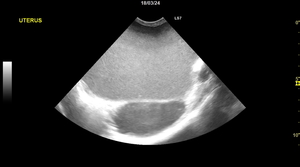
・Prostate (prostate hypertrophy) ・Uterus (uterine edema)
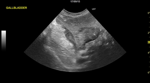
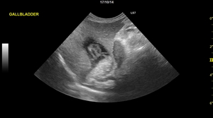
・Gall bladder (cholelithiasis) ・Gall bladder (gall bladder rupture)
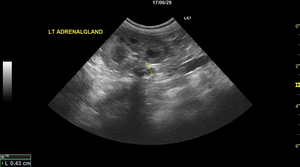
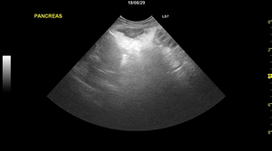
・Adrenal gland ・Pancreas (pancreatitis)
◆EYES ★uses a special eye examination probe
・Cataract ・Vitreous inflammation
・Retinal detachment
……etc.